Global / English
A Highly Photostable Fluorescent Protein
- Technology That Allows For Quantitative Observation of Cellular Microstructures and Viruses -
A joint research group consisting of researchers from the Laboratory for Cell Function Dynamics of the RIKEN Center for Brain Science and the Biotechnological Optics Research Team of the RIKEN Center for Advanced Photonics; the Asamushi Research Center for Marine Biology, Graduate School of Life Sciences, Tohoku University; the Ōmura Satoshi Memorial Institute, Kitasato University; and the Safety Science Research Laboratories, Kao Corporation, promotes sustainable bioimaging by utilizing a jellyfish-derived green fluorescent protein StayGold, which is bright and exhibits minimal fading. StayGold improves spatiotemporal resolution and dramatically extends the observation period. This finding offers a new and powerful tool for the bioimaging community, in which most experimenters encounter the challenging issue of photobleaching[1] of fluorescent proteins.
On the basis of gene expression[2] analysis of the jellyfish Cytaeis uchidae[3] done by researchers from Tohoku University, researchers from RIKEN created StayGold, a bright fluorescent protein variant with the unique property of minimal photobleaching, through their molecular cloning and mutagenesis studies on a wild-type fluorescent protein. Fluorescent labelling of organelles such as the endoplasmic reticulum, mitochondria, and microtubules with StayGold has revealed dynamic structural changes that could not be observed previously due to the photobleaching of conventional fluorescent proteins. In addition, StayGold was molecularly linked to the anti-Spike VHH antibody[4] developed by researchers from Kitasato University and Kao Corporation to visualize the process of virion maturation in SARS-CoV-2-infected cells, which is appealing to scientists in medicine who are working furiously to develop ways to reliably detect SARS-CoV-2 and other infectious viruses.
These findings were reported in Nature Biotechnology Online (published on April 25).
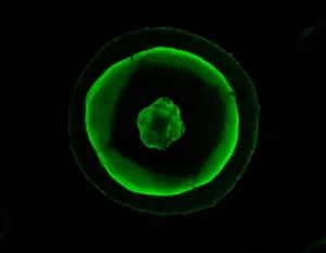
Green fluorescence emitted from the jellyfish Cytaeis uchidae
(Top view of jellyfish umbrella imaged under blue light. The diameter of the umbrella is 1.3 mm.)
Publication information
Title:
A highly photostable and bright green fluorescent protein
Authors:
Masahiko Hirano1, Ryoko Ando2, Satoshi Shimozono2, Mayu Sugiyama2, Noriyo Takeda3,10,
Hiroshi Kurokawa2, Ryusaku Deguchi4, Kazuki Endo4,11, Kei Haga5, Reiko Takai-Todaka5, Shunsuke Inaura6, Yuta Matsumura6, Hiroshi Hama2, Yasushi Okada 7,8, Takahiro Fujiwara9, Takuya Morimoto6, Kazuhiko Katayama5, and Atsushi Miyawaki1,2
1Biotechnological Optics Research Team, RIKEN Center for Advanced Photonics, Saitama, Japan. 2Laboratory for Cell Function Dynamics, RIKEN Center for Brain Science, Saitama, Japan. 3Asamushi Research Center for Marine Biology, Tohoku University, Aomori, Japan. 4Department of Biology, Miyagi University of Education, Sendai, Japan. 5Department of Infection Control and Immunology, Ōmura Satoshi Memorial Institute, Kitasato University, Tokyo, Japan. 6Safety Science Laboratories, Kao Corporation, Tokyo, Japan. 7Laboratory for Cell Polarity Regulation, RIKEN Center for Biosystems Dynamics Research, Osaka, Japan. 8Department of Cell Biology and Department of Physics, UBI and WPI-IRCN, The University of Tokyo, Tokyo, Japan. 9Institute for Integrated Cell-Material Sciences, Kyoto University, Kyoto, Japan. 10Current address: Graduate School of Integrated Sciences for Life, Hiroshima University, Hiroshima, Japan. 11Current address: Narita Elementary School, Miyagi, Japan.
Journal:
Nature Biotechnology
DOI:
10.1038/s41587-022-01278-2
Footnote
[1] Photobleaching
A chromophore is a structural unit that absorbs the visible range of light and determines the color of objects. Fluorescent proteins form their chromophores internally. Degradation of chromophores causes photobleaching, leading to irreversible loss of color. This loss of color inevitably results in loss of fluorescence.
[2] Gene expression analysis
In the context of this article, this refers to data obtained from comprehensive analyses (transcriptome analyses) of messenger RNA (mRNA) of particular tissues.
[3] Cytaeis uchidae
The Cytaeis uchidae is a jellyfish belonging to the Cnidaria division, Hydrozoa class, and Anthomedusae order. Individual Cytaeis uchidae have a spherical body 1–2 mm in diameter. Cytaeis uchidae can be raised in laboratories and has drawn attention as an experimental material for reproductive biology and a school science material.
[4] VHH antibody
Camelids such as alpacas produce antibodies formed only of heavy chains with no light chains. The variable domain of this heavy chain antibody is referred to as VHH (variable domain of heavy chain of heavy chain) and is used as the smallest protein segment that recognizes antigens. Haga et al. (Kitasato University) have demonstrated that symptoms of SARS-CoV-2 infected hamsters could be alleviated using the anti-SARS-CoV-2 spike VHH antibody.