Global / English
Skin Science for Development of Cosmetics
Dermatological science
Our dermatological research has been evolving day after day, thankfully attributed to long-term, persistent studies and cutting-edge technological development.
Kao has been studying “hyaluronic acid” in the skin as a pioneer and front-runner for over 40 years*1–5 . A major part of hyaluronic acid produced in the human body exists in the skin and has various physiological functions: not only contributes to hydration of the skin, but also cell proliferation, differentiation, and migration.
We were the first worldwide to shed light on the molecular mechanisms of the synthesis and degradation of hyaluronic acid in the skin, and revealed the relationship between their changes and photoaging symptoms (skin wrinkles and sagging). Furthermore, we have developed the original and high-quality cosmetic materials based on our unique concept “hyaluronic acid from within the skin” by maintaining the balance between the synthesis and degradation of hyaluronic acid.
-
* 1 Sugiyama, Y., Shimada, A., Sayo, T., Sakai, S., Inoue, S. (1998). Putative hyaluronan synthase mRNA are expressed in mouse skin and TGF-β upregulates their expression in cultured human skin cells. Journal of Investigative Dermatology, 110(2), 116-121.
-
* 2 Sayo, T., Sugiyama, Y., Takahashi, Y., Ozawa, N., Sakai, S., Inoue, S., Ishikawa, O., Tamura, M. (2013). Hyaluronan synthase 3 regulates hyaluronan synthesis in cultured human keratinocytes. Journal of Investigative Dermatology, 118(1), 43-48.
-
* 3 Yoshida, H., Nagaoka, A., Kusaka-Kikushima, A., Tobiishi, M., Kawabata, K., Sayo, T., Sakai, S., Sugiyama, Y., Enomoto, H., Okada, Y., Inoue, S. (2013). KIAA1199, a deafness gene of unknown function, is a new hyaluronan binding protein involved in hyaluronan depolymerization. Proceedings of the National Academy of Sciences of the United States of America, 110(14), 5612-5617.
-
* 4 Yoshida H., Nagaoka A., Komiya A., Aoki M., Nakamura S., Morikawa T., Ohtsuki R., Sayo T., Okada Y., Takahashi Y. (2018). Reduction of hyaluronan and increased expression of HYBID (alias CEMIP and KIAA1199) correlate with clinical symptoms in photoaged skin. British Journal of Dermatology. 179(1), 136-144.
-
* 5 Endo, Y., Yoshida, H., Akazawa, Y., Yamazaki, K., Ota, Y., Sayo, T., Takahashi, Y. (2022). Antiwrinkle efficacy of 1-ethyl-β-N-acetylglucosaminide, an inducer of epidermal hyaluronan production. Skin Research & Technology, 28(1), 58-65.
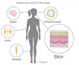
Distribution of hyaluronic acid in the body
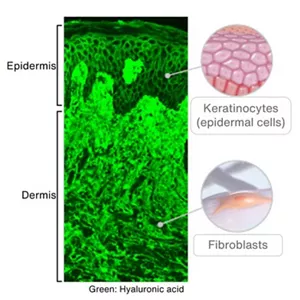
Image of hyaluronic acid in the skin
The skin has a complex, multi-layer structure consisting of the epidermis and dermis, in addition to several structural materials including collagen and blood vessels. Kao has developed CT-Skin®, which employs optical interferometric information to create digital 3D images of the inner skin. CT-Skin® enables us to visualize static conditions of the inner skin non-invasively, as well as dynamic changes in the skin by observing the same site over time.
We will continue to deepen our understanding of the functioning of the skin and its mechanisms to develop attractive cosmetics based on scientific evidence by applying state-of-the-art technologies.
- Home
- Innovation
- Research & Development
- Fundamental Research
- Biological Science
- Skin Science for Development of Cosmetics
- Home
- Innovation
- Research & Development
- Fundamental Research
- Biological Science
- Skin Science for Development of Cosmetics